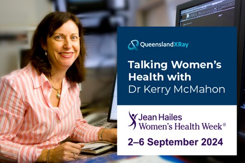
Women’s Health Week, 2nd – 6th September, is a week dedicated to shining a light on some of the biggest issues in women’s health today and led by not-for-profit Jean Hailes for Women’s Health. Throughout this week we will be showcasing some of our Radiologists who specialise in women’s imaging, who share their perspectives on what it means to work in women’s health.
We were also fortunate enough to grab some of Dr McMahon’s time who shared her thoughts about women’s healthcare in Australia.
Dr McMahon enjoys working in women’s imaging as it is an opportunity to work in a great team, with tremendous clinical interaction with patients, many of whom are seen on an annual basis over many years. She says that “women’s imaging incorporates all aspects of caring for a woman – from conception in the womb, to adolescent issues and menstrual disorders, to fertility assessment and pregnancy, as well as later in life through breast imaging and Bone Mineral Densitometry in older women”.
Today, Australian women have access to unprecedented medical information to assist them in looking after their health. The last 10-20 years have seen tremendous technological developments occur in the area of breast imaging which has had a positive impact for the early detection of disease. Dr McMahon reflects on when breast MRI was introduced in Australia when she first started working with Queensland X-Ray. “In those first few years we used to see patients fly in from all over Australia to have a breast MRI with Queensland X-Ray as we were the first centre in Australia to establish this modality. Now, this technology is common across all regional areas. Digitisation of mammography was late (only really occurring in the last 10 years) and now 3D digital tomosynthesis is routine. We are now moving to 3D breast densitometry and risk assessment ad contrast mammography in the near future and this will improve thing even further.
Australia has one of the best health systems in the world. However, that being said, access to healthcare can be more accessible for some more than others. When contemplating this issue, Dr McMahon tells us that “although we work in private practice, the vast majority of our patients are not affluent, are generally the regular mums and dads trying hard to make ends meet, and do not have private health insurance”. Nearly all early first trimester pregnancy assessments are performed and triaged through private radiology with the GP, as are most breast lumps. As such, the radiologist works very closely with referring GP’s in sorting out initial problems and with the management of pregnancy. By way of example, Dr McMahon explains that “a patient may be referred from their GP with pelvic pain in early pregnancy and ultrasound may detect an ectopic pregnancy. We discuss these findings directly with the patient and the referring doctor and arrange transfer of the patient to an emergency centre for appropriate management.” Queensland X-Ray makes access to services as equitable as possible by keeping gap fees affordable. Queensland X-Ray also provides services to those who do not have the means or are on a health care card/pensions without the need to pay a gap.
Dr McMahon enjoys the opportunity to see so many patients on a regular basis and getting to know them personally. Over many years she has seen numerous patients through various difficult journeys. Here, she describes a series of interactions with one patient in particular who encountered many challenges. Dr McMahon saw her over a period of approximately 10 years in relation to a number of issues ranging from difficulty falling pregnant to further tragic news.
“I will always remember this patient for the resilience she showed in the face of adversity. Sadly, after many attempts and finally receiving a positive pregnancy test, her ultrasound showed an ectopic pregnancy which was devastating to her. Fortunately, some months later she finally fell pregnant again. She presented at 12 weeks for her nuchal scan, extremely excited and happy, however the most difficult of situations had occurred. When the probe was placed on her belly there was no heartbeat and it was confirmed the baby had died. Somehow she coped with this news and fell pregnant for a third time. She had a normal 12-week scan and morphology study. However, later in the pregnancy she had an unusual thickening of her breast. She decided to attend the QEII Hospital practice for this scan because she specifically did not want to see me at Mater Women’s (as I was so often the bearer of bad news!). It was crazy, but I happened to be the doctor on at QEII that day – such a rare thing these days. Unfortunately the ultrasound was not normal – she had a diffusely infiltrative lobular carcinoma (a type of breast cancer). I saw her yearly for the next five years and she progressed well through treatment. During this time, she suffered enormous life challenges including the loss of her partner. Then, 6 years later when her daughter had just started school, she came for a routine check. She was extremely excited as she had recently met a new partner and thought this was the beginning of a new chapter in her life. Unfortunately her imaging was not good; we had found recurrence. Tragically, she died just 6 months later, her beautiful child who I had seen since conception was now an orphan. Sally Graham (the breast care nurse) and so many of the Queensland X-Ray staff all saw her through so many poignant aspects of her life. Whilst this case ended in tragedy, there are fortunately may patients with good outcomes and good result. It is wonderful working with so many dedicated people and there are many clerical staff, sonographers and mammographers in our department who have been seeing the same patients for over 20 years! We all love this aspect of the job”.

